
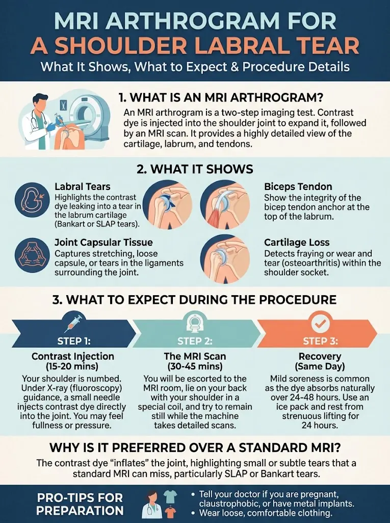
Shoulder pain that worsens when you reach overhead, lift weights, or throw a ball can make everyday activities uncomfortable. If your clinician suspects a labral tear, they may recommend an MRI arthrogram to examine the cartilage-like tissue around your shoulder socket and look for damage that a routine examination cannot confirm.
An MRI arthrogram can provide detailed images of the shoulder labrum, but it does not automatically prove that a tear is causing your symptoms. Some labral changes appear on scans in people who have little or no shoulder pain, so imaging findings must be interpreted alongside your symptoms, physical examination, and activity history.
This guide explains how an MRI arthrogram for a shoulder labral tear works, why contrast is used, what the scan may reveal, how it compares with a standard MRI, and what happens after the results. Understanding these details can help you prepare for the test and have a more informed discussion with your healthcare professional.
What Is an MRI Arthrogram for a Shoulder Labral Tear?
An MRI arthrogram is an imaging test that combines magnetic resonance imaging (MRI) with contrast fluid injected into the shoulder joint. The fluid expands the joint space and can make certain structures, including the labrum, easier to assess.
The labrum is a ring of firm fibrocartilage that deepens the shoulder socket and helps support joint stability. A tear may occur in different areas. A SLAP tear, for example, affects the upper labrum from front to back and may involve the attachment of the biceps tendon.
To understand the broader range of injuries, see the different types of shoulder labral tears.
What Can an MRI Arthrogram Show?
An MRI arthrogram may help identify the location and extent of a labral tear, whether the tear extends into nearby structures, and whether other abnormalities could contribute to symptoms. It may also help clinicians assess the biceps tendon attachment and other soft tissues around the shoulder.
| Finding | What it may indicate | Important limitation |
|---|---|---|
| Contrast tracking into or beneath the labrum | A possible labral tear or separation | Interpretation depends on the tear location and normal anatomical variations |
| Irregular or detached labral tissue | Possible structural damage | The scan alone cannot establish how much pain it causes |
| Changes near the biceps anchor | Possible involvement of the biceps-labrum attachment | Symptoms and examination findings still matter |
| Other soft-tissue abnormalities | Possible additional causes of shoulder pain | Some findings may be incidental |
The report may describe a tear’s location, size, or associated changes, but the wording can be difficult to interpret without clinical context.
For a closer look at standard imaging, read what a shoulder labral tear looks like on MRI.
MRI Arthrogram vs. Standard Shoulder MRI
Both tests use magnetic fields to produce detailed images without ionizing radiation. The key difference is that an arthrogram involves placing contrast directly inside the shoulder joint before the MRI.
| Feature | Standard MRI | MRI arthrogram |
|---|---|---|
| Contrast placement | Usually no contrast inside the joint | Contrast is injected into the joint |
| Labrum assessment | Can identify many labral abnormalities | May better outline selected tears or subtle separations |
| Invasiveness | No joint injection is usually required | Requires an injection and carries additional risks |
| Best choice | Depends on the suspected problem and local imaging protocol | Considered when more detailed joint assessment may help |
An arthrogram is not automatically necessary for every shoulder injury. The choice depends on the suspected diagnosis, the initial examination, prior imaging, and whether the additional detail is likely to change treatment. You can also explore how a labral tear differs from a rotator cuff tear and how a labral tear differs from shoulder impingement.
What Happens During an MRI Arthrogram?
The process generally involves two stages: placing the contrast fluid and taking the MRI images.
- Preparation: The imaging team reviews your medical history, implants, allergies, medications, and other safety considerations. Tell the team if you may be pregnant or have had a previous reaction to contrast or an injection.
- Joint injection: A trained clinician cleans the skin and guides a needle into the shoulder joint, sometimes using imaging guidance. A small amount of contrast fluid is injected.
- MRI scan: You lie still while the scanner captures images. You may hear loud tapping or knocking sounds, and the scan can take several minutes or longer depending on the protocol.
- Review: A radiologist interprets the images and sends a report to the clinician who ordered the test.
The injection may cause pressure, temporary soreness, or discomfort. The MRI itself should not hurt, although holding the shoulder in one position can be uncomfortable if it is already painful.
Does the Injection Hurt, and What Are the Risks?
People experience the injection differently. Local anesthetic may be used to reduce discomfort, but you may still feel a brief sting, pressure, or fullness in the joint.
Temporary soreness after the procedure can occur. Less commonly, complications include bleeding, infection, or a reaction to the injected material. Tell the imaging team about relevant allergies, bleeding disorders, immune-system conditions, and medications that may affect bleeding; do not stop prescribed medication unless your clinician instructs you to.
Contact a healthcare professional promptly if you develop increasing pain, spreading redness, warmth, fever, or significant swelling after the injection.
How Accurate Is an MRI Arthrogram?
An MRI arthrogram can improve visualization of certain labral abnormalities, but no imaging test is perfect. Small tears, normal anatomical variations, and differences in scanning technique or interpretation can affect the result.
A scan also cannot determine by itself whether a finding is the main source of pain. For example, a person may have a labral abnormality alongside rotator cuff irritation, tendon problems, or shoulder stiffness. Your clinician will compare the report with early symptoms of a shoulder labral tear and examination findings before deciding what the result means.
What Happens If the MRI Arthrogram Shows a Tear?
A tear on the report does not automatically mean you need surgery. Treatment depends on symptoms, the type and location of the tear, shoulder stability, activity demands, and whether other conditions are present.
| Result and clinical picture | Possible next step |
|---|---|
| Tear or abnormality with manageable symptoms | Activity modification and a clinician-guided rehabilitation plan |
| Persistent pain or reduced function | Further evaluation and targeted physical therapy |
| Recurrent instability or significant symptoms despite treatment | Specialist assessment to discuss additional options |
| Imaging finding that does not match symptoms | Reassessment for another or additional cause |
Non-surgical care may include modifying painful overhead activities, appropriate pain relief, and physical therapy to improve shoulder movement, rotator cuff strength, and shoulder-blade control. Ask your clinician which movements are appropriate; avoid pushing through sharp pain or a sense that the shoulder is slipping.
For a detailed comparison, see shoulder labral tear treatment: physical therapy or surgery. If rehabilitation is recommended, physical therapy exercises and recovery for a SLAP tear can help you understand the general approach. Avoid choosing exercises without considering your particular injury; exercises to avoid with a shoulder labral tear may be useful to discuss with your therapist.
When Should You Seek Medical Care?
Arrange a medical evaluation for persistent shoulder pain, painful clicking or catching, weakness, or difficulty with overhead tasks, particularly when symptoms began after an injury. Seek urgent care after a major injury if the shoulder looks deformed, you cannot move the arm, or you develop new severe weakness, numbness, or a pale or cold hand. Fever with a hot, swollen shoulder—especially after an injection—also requires prompt assessment.
While waiting for evaluation, avoid activities that reliably trigger pain, and support the arm in a comfortable position during rest. For nighttime discomfort, sleeping positions that may help with a SLAP tear may offer practical ideas.
Frequently Asked Questions
1. Is an MRI arthrogram better than a regular MRI for a labral tear?
It may provide extra detail for selected labral injuries, but it is not always necessary. Your clinician will consider the suspected injury and whether the additional information could affect treatment.
2. Can an MRI arthrogram miss a shoulder labral tear?
Yes. Imaging has limitations, and small or subtle abnormalities may not be clearly identified. Persistent symptoms may warrant reassessment even if the report does not confirm a tear.
3. How long does a shoulder MRI arthrogram take?
The full appointment includes preparation, the joint injection, and MRI imaging, so it takes longer than the scan alone. Timing varies by facility and imaging protocol.
4. Can I use my arm after the procedure?
You can often resume light activities as tolerated, but temporary soreness may occur. Follow the imaging team’s instructions and avoid strenuous shoulder activity if it worsens discomfort.
5. Does contrast from an MRI arthrogram stay in the shoulder?
The injected fluid is gradually absorbed by the body. Your care team can explain the contrast used and any precautions relevant to your medical history.
6. Does a labral tear on MRI always cause pain?
No. Some labral changes may be present without symptoms, especially as people get older. Clinicians interpret the images alongside the location of your pain, examination, and activity history.
7. Will I need surgery if the arthrogram shows a SLAP tear?
Not necessarily. Many people begin with activity modification and rehabilitation, while surgery may be considered for selected cases when symptoms persist or other factors support it.
8. Can an X-ray show a shoulder labral tear?
An X-ray does not directly show the labrum because it primarily shows bones. It can still help identify fractures, arthritis, or other bony changes that may be relevant.
9. Is an MRI arthrogram safe if I have metal implants?
Some implants are MRI-compatible and others require special precautions or may make MRI unsafe. Provide the imaging team with the exact implant details before the appointment.
10. What should I ask my doctor after the results?
Ask where the abnormality is, whether it matches your symptoms, whether other structures are involved, and what treatment options are reasonable. You can also discuss how SLAP tears are classified and compared with Bankart tears if the report uses unfamiliar terminology.
Conclusion
An MRI arthrogram can help clinicians evaluate a suspected shoulder labral tear by outlining joint structures in greater detail. The result is only one part of the assessment: symptoms, examination findings, activity needs, and other possible causes of pain all matter. Ask your clinician how the findings relate to your symptoms and whether rehabilitation, further evaluation, or another treatment approach is appropriate.
Disclaimer: This article is provided for educational and informational purposes only.
This article is for education purpose only and is not a diagnosis or a substitute for medical advice.