
A burning, electric, or pins-and-needles sensation that seems to live inside the tissue rather than on the surface. It often arrives without obvious provocation, worsens with certain positions or activities, and can be maddeningly difficult to pinpoint. Whether it appears in the wrist and hand or the ankle and foot, this type of pain signals that a nerve is being squeezed somewhere along its pathway, deprived of space, blood supply, or both.
Carpal tunnel syndrome is well known — most people have heard of it, and it is among the most commonly diagnosed peripheral nerve conditions in the world. Tarsal tunnel syndrome is its lesser-known counterpart in the foot, following the same mechanical principle but in a different anatomical location. Because both conditions share a similar pathological mechanism and symptom profile, they are often confused or misdiagnosed — and because tarsal tunnel syndrome is less familiar even to many clinicians, it can go unrecognized for months or years. Understanding the differences — and the similarities — between these two conditions is essential for getting the right diagnosis and the right treatment.
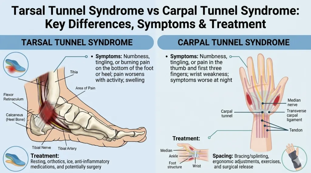
Tarsal tunnel syndrome involves compression of the posterior tibial nerve as it passes through the tarsal tunnel, a narrow fibrous canal on the inner side of the ankle. Carpal tunnel syndrome involves compression of the median nerve as it passes through the carpal tunnel, a narrow channel in the wrist formed by the carpal bones and the transverse carpal ligament. Both tunnels are unyielding anatomical structures — any increase in the contents within them, or any thickening of the surrounding tissue, reduces the available space and compresses the nerve. This shared principle is where the similarities are most striking — and where an understanding of one condition illuminates the other.
The Anatomy Behind the Conditions
The Carpal Tunnel
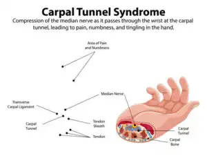
The carpal tunnel is a narrow passageway at the base of the palm, bounded on three sides by the carpal bones and on the fourth by the transverse carpal ligament. Through this tunnel passes the median nerve — which supplies sensation to the thumb, index finger, middle finger, and the thumb side of the ring finger — along with nine flexor tendons that bend the fingers. When the contents of this tunnel swell or the ligament thickens, the median nerve is compressed, producing the characteristic symptoms of carpal tunnel syndrome.
The Tarsal Tunnel
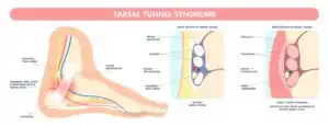
The tarsal tunnel runs along the inner (medial) side of the ankle, just behind the medial malleolus (the bony bump on the inside of the ankle). It is bounded by the ankle bones on one side and the flexor retinaculum — a tough band of fibrous tissue — on the other. Through this tunnel pass the posterior tibial nerve, the posterior tibial tendon, and the blood vessels supplying the foot. The posterior tibial nerve divides within or just below the tarsal tunnel into branches that supply sensation to the heel, arch, and sole of the foot and motor function to the intrinsic muscles of the foot.
For a broader understanding of how nerve compression in the lower limb relates to other conditions, our guide on heel pain when walking causes explores the overlap between tarsal tunnel and plantar heel pain in depth.
Comparing Symptoms: What’s the Same, What’s Different?
Carpal Tunnel Syndrome Symptoms
Carpal tunnel syndrome typically presents with tingling, numbness, and burning pain in the thumb, index, middle, and part of the ring finger. The little finger is notably spared, as it is supplied by the ulnar nerve which does not pass through the carpal tunnel. Symptoms are often worse at night and during activities that involve sustained wrist flexion or extension — such as typing, driving, or holding a phone.
Characteristic features of CTS:
- Nocturnal tingling and numbness that wakes patients from sleep, often relieved by shaking or “flicking” the hand (the “flick sign”).
- Weakness and clumsiness of grip, particularly for fine motor tasks like buttoning clothing or handling small objects.
- Pain that may radiate up the forearm toward the elbow.
- Wasting (atrophy) of the thenar muscles at the base of the thumb in advanced or long-standing cases.
- Positive Phalen’s test: reproduction of symptoms when the wrist is held in flexion for 60 seconds.
- Positive Tinel’s sign: tingling produced by tapping over the carpal tunnel at the wrist.
For those interested in how early nerve compression signs present, our article on early signs of carpal tunnel syndrome covers the subtle initial presentations that precede the classic symptom picture.
Tarsal Tunnel Syndrome Symptoms
Tarsal tunnel syndrome produces pain, burning, tingling, and numbness along the inner ankle, heel, and sole of the foot. Symptoms are often described as electric, hot, or shooting in quality and may radiate into the arch and toes. The location of pain depends on which branch of the posterior tibial nerve is most compressed. Unlike plantar fasciitis, which typically causes pain specifically at the heel attachment point and is worst in the morning, tarsal tunnel pain is more diffuse, extends along the nerve distribution, and often worsens with prolonged standing or walking.
Characteristic features of TTS:
- Burning, tingling, or electrical pain on the inner ankle and sole of the foot, worsened by prolonged weight-bearing.
- Pain that may continue at rest or at night, though typically less intense than during activity.
- Positive Tinel’s sign at the tarsal tunnel: tapping behind the medial malleolus reproduces symptoms.
- Weakness of the intrinsic foot muscles in advanced cases, manifesting as difficulty with toe movements.
- Symptoms may be aggravated by foot positions that increase tension in the tarsal tunnel — such as excessive pronation, which is why flat feet are a significant risk factor. Our detailed guide on flat feet symptoms and treatment explains this biomechanical relationship.
Head-to-Head Comparison
| Feature | Carpal Tunnel Syndrome | Tarsal Tunnel Syndrome |
| Nerve compressed | Median nerve | Posterior tibial nerve |
| Location | Wrist | Inner ankle |
| Pain location | Hand, fingers (thumb to ring finger) | Foot, heel, sole |
| Night symptoms | Very common | Present but less predominant |
| Worsened by | Wrist flexion, prolonged grip | Prolonged standing, walking, pronation |
| Sensory loss area | Thumb, index, middle, ring fingers | Heel, arch, sole |
| Motor weakness | Thenar muscles (thumb) | Intrinsic foot muscles |
| Diagnostic test | Nerve conduction studies, Phalen’s, Tinel’s | Nerve conduction studies, Tinel’s at ankle |
| Common associations | Pregnancy, diabetes, thyroid, repetitive wrist use | Flat feet, ankle trauma, diabetes, varicose veins |
Causes and Risk Factors
Shared Risk Factors
Both conditions share several systemic risk factors:
Systemic conditions that increase risk for both:
- Diabetes mellitus, which causes peripheral nerve vulnerability through diabetic neuropathy, making nerves more susceptible to compression injury.
- Hypothyroidism, which can cause tissue swelling and fluid retention within confined anatomical tunnels.
- Inflammatory conditions such as rheumatoid arthritis, which produce synovitis that reduces tunnel space. Our comparison of rheumatoid arthritis vs osteoarthritis explains how inflammatory joint disease affects surrounding soft tissue structures.
- Pregnancy, where hormonal changes increase ligament laxity and fluid retention, compressing both tunnels.
- Obesity, which increases tissue bulk and systemic inflammation.
Causes Specific to Carpal Tunnel Syndrome
Repetitive hand and wrist use — particularly sustained keyboard work, assembly line tasks, and use of vibrating tools — is among the most important occupational risk factors for CTS. A direct traumatic wrist injury such as a Colles’ fracture can alter the geometry of the carpal tunnel and predispose to nerve compression during healing. Ganglion cysts or tumours within the tunnel can occupy space and directly compress the nerve.
Causes Specific to Tarsal Tunnel Syndrome
Flat feet and overpronation are the most significant biomechanical contributors to TTS. The inward rolling of the foot stretches and compresses the tarsal tunnel contents with each step. Prior ankle sprains or fractures can cause scar tissue formation within the tunnel. Space-occupying lesions such as lipomas, ganglion cysts, or varicose veins within the tarsal tunnel can directly compress the posterior tibial nerve. Heel pain conditions that cause localised inflammation near the tarsal tunnel — including the conditions discussed in our guide on heel pain when walking causes — may contribute to secondary nerve irritation.
Diagnosis
How Carpal Tunnel Syndrome is Diagnosed
Clinical diagnosis of CTS relies on the characteristic symptom distribution and positive provocative tests (Phalen’s and Tinel’s). Nerve conduction studies (NCS) and electromyography (EMG) are the gold-standard investigations, objectively measuring the degree of slowing across the carpal tunnel and confirming median nerve compromise. Ultrasound has emerged as a valuable additional tool, allowing direct visualisation of median nerve swelling and cross-sectional area.
How Tarsal Tunnel Syndrome is Diagnosed
TTS diagnosis is more challenging because the symptom pattern overlaps with several other common foot conditions — plantar fasciitis, peripheral neuropathy, and lumbar radiculopathy (sciatica). Our article on sciatica causes, symptoms and treatment clarifies the distinction between lumbar nerve root pain and peripheral nerve entrapment, which is clinically important. Nerve conduction studies across the tarsal tunnel, combined with EMG of the intrinsic foot muscles, provide objective confirmation. MRI is particularly valuable in TTS for identifying space-occupying lesions within or around the tarsal tunnel that may be amenable to surgical decompression.
Treatment Options: Parallels and Differences
Non-Surgical Treatment
Both conditions respond to a broadly similar conservative management framework, though the specific modalities differ based on anatomy:
Conservative treatments for CTS:
- Wrist splinting in a neutral position — particularly at night — reduces carpal tunnel pressure during wrist flexion and is the most effective initial intervention.
- Activity modification to reduce repetitive wrist loading.
- Corticosteroid injections into the carpal tunnel provide reliable short-term relief and are often used as a bridge to surgery or a trial of conservative management.
- Ergonomic keyboard and workstation adjustments to reduce sustained wrist loading.
- Specific tendon and nerve gliding exercises to reduce adhesions and improve nerve mobility within the tunnel.
Conservative treatments for TTS:
- Foot orthotic insoles and motion-control footwear to correct overpronation and reduce dynamic compression of the tarsal tunnel.
- Ankle bracing or taping to control excessive hindfoot pronation.
- Rest and activity modification to reduce neural load.
- Anti-inflammatory medication for acute flare-ups.
- Corticosteroid injection into the tarsal tunnel to reduce local inflammation and tunnel pressure.
- Management of any underlying systemic condition contributing to nerve vulnerability.
For those interested in how lifestyle factors — including diet and weight — influence nerve and joint conditions, our article on how changing your diet can help reduce pain naturally offers evidence-based dietary strategies that apply broadly to inflammatory nerve conditions.
Surgical Treatment
Surgery is considered when conservative management has failed and the condition significantly impairs daily function or quality of life.
Carpal tunnel release is one of the most commonly performed surgical procedures worldwide. It involves dividing the transverse carpal ligament to decompress the median nerve. It can be performed as an open procedure or endoscopically. Results are excellent, with high rates of symptom resolution and return to full function. Recovery is typically complete within 4–6 weeks.
Tarsal tunnel release involves dividing the flexor retinaculum to decompress the posterior tibial nerve and its branches. Surgical outcomes for TTS are less predictably excellent than for CTS, largely because the underlying cause is more variable. When a discrete compressive lesion (such as a cyst or ganglion) is identified and removed, outcomes are generally very good. When compression is due to diffuse scarring or ongoing biomechanical factors, results can be less consistent.
Recovery and Prognosis
For carpal tunnel syndrome, non-surgical management provides long-term relief in approximately 60–70% of mild to moderate cases. Surgical decompression produces symptom relief in 85–95% of patients, with motor function recovering more slowly than sensory symptoms. Delay in treatment allows progressive nerve damage that may not be fully reversible.
For tarsal tunnel syndrome, outcomes depend heavily on identifying and addressing the primary cause. Cases related to a treatable structural cause — a cyst, varicose vein, or scar tissue — have the best surgical prognosis. Cases driven by ongoing biomechanical issues such as flat feet require concurrent correction of foot mechanics to achieve lasting relief.
In both conditions, the longer compression persists and the more severe the nerve damage, the less complete the recovery. This underscores the importance of early diagnosis and intervention. For context on how other musculoskeletal conditions are managed with a view to protecting long-term function, our article on cervical spondylosis symptoms causes and treatment explores a similar principle of early intervention for nerve-related spinal conditions.
Prevention Tips
Preventing or reducing the risk of both conditions:
- Manage systemic health conditions — particularly diabetes, thyroid disorders, and excess weight — that increase nerve vulnerability throughout the body.
- Maintain healthy body weight to reduce soft-tissue bulk and systemic inflammatory load on nerve passages.
- For CTS: optimise workstation ergonomics, take regular movement breaks from keyboard work, and use voice recognition or ergonomic input devices if repetitive wrist strain is unavoidable.
- For TTS: wear supportive footwear with motion control, address flat feet proactively with orthotics and targeted exercises, and protect the ankle from repeated sprains with appropriate footwear and training load management.
Conclusion
Tarsal tunnel syndrome and carpal tunnel syndrome are both peripheral nerve entrapment conditions sharing the same fundamental mechanism — a nerve compressed within a confined anatomical tunnel. They share risk factors, diagnostic principles, and a broadly parallel treatment philosophy. The key differences lie in their location, the specific nerve involved, and the biomechanical factors that drive them. Both conditions respond well to early, appropriate management, and both can lead to permanent nerve damage if left untreated. Whether the burning, tingling sensation lives in your hand or your foot, the message is the same: get an accurate diagnosis, address the cause, and begin treatment before the nerve has suffered irreversible damage.
Frequently Asked Questions
- Can you have both carpal tunnel and tarsal tunnel syndrome at the same time? Yes. People with systemic conditions such as diabetes or hypothyroidism are at elevated risk for multiple peripheral nerve entrapments simultaneously. This is sometimes referred to as “double crush” syndrome when two entrapment points affect the same nerve pathway.
- How do I know if I have tarsal tunnel syndrome or plantar fasciitis? Plantar fasciitis causes pain specifically at the heel insertion point, is worst with the first steps of the morning, and improves with activity. Tarsal tunnel syndrome produces burning, tingling, or electric pain along the inner ankle and sole, can occur at rest or at night, and is reproduced by tapping over the tarsal tunnel behind the medial malleolus.
- Is carpal tunnel syndrome permanent? Without treatment, progressive nerve damage can cause permanent sensory loss and muscle wasting. With timely appropriate treatment — particularly surgical release in moderate to severe cases — the majority of patients recover full or near-full function.
- Can flat feet cause tarsal tunnel syndrome? Yes. Flat feet and overpronation stretch the tarsal tunnel with each step, increasing pressure on the posterior tibial nerve. Correcting foot mechanics with orthotics and footwear is an important component of tarsal tunnel management.
- What makes carpal tunnel worse at night? During sleep, many people naturally flex their wrists, increasing pressure within the carpal tunnel. The supine position may also contribute to fluid redistribution, slightly increasing tissue pressure within the tunnel.
- How long does carpal tunnel surgery take to recover from? Most patients return to light activities within 1–2 weeks. Full recovery, including resolution of nerve symptoms and return to heavy manual work, typically takes 4–12 weeks. Nerve regeneration after severe compression can continue for 6–12 months.
- What nerve conduction tests are used for diagnosis? Nerve conduction velocity (NCV) and electromyography (EMG) measure the speed and efficiency of nerve signals across the compressed segment. In CTS, slowing across the wrist is the key finding; in TTS, slowing across the tarsal tunnel. These tests also help grade severity and guide treatment decisions.
- Are there exercises to help tarsal tunnel syndrome? Neural mobilisation (nerve gliding) exercises for the tibial nerve, arch strengthening exercises, and calf stretching can reduce tension and improve nerve mobility. These should be performed gently within a pain-free range under physiotherapist supervision.
- Can tarsal tunnel syndrome resolve without surgery? Many cases respond to conservative management including orthotics, footwear modification, activity adjustment, and injection therapy. Surgery is reserved for cases that fail to respond to sustained conservative treatment and is more clearly indicated when a discrete compressive lesion is identified.
- What is the difference between tarsal tunnel syndrome and peripheral neuropathy? Peripheral neuropathy causes generalised, symmetric sensory symptoms — typically a “stocking and glove” distribution — and is caused by systemic nerve disease (most commonly diabetes). Tarsal tunnel syndrome causes localised symptoms in the specific distribution of the posterior tibial nerve, on one or both sides, and is caused by local nerve compression rather than generalised nerve disease.
Medical Disclaimer
The information in this article is for educational purposes and does not constitute professional medical advice, diagnosis, or treatment.
This article is for education purpose only and is not a diagnosis or a substitute for medical advice.