
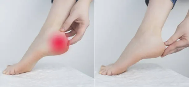
That sharp, stabbing pain in your heel the moment your foot hits the floor in the morning — or the dull, grinding ache that builds with every step on a long walk — is one of the most disruptive discomforts a person can experience. Heel pain when walking doesn’t just slow you down; it changes the way you move, stand, and go about your entire day. Many people begin unconsciously favouring one foot, altering their gait, and putting stress on their knees, hips, and lower back as a secondary consequence. Over time, what begins as a minor foot problem can quietly ripple into a full-body postural issue.
If you’ve been dealing with heel pain, you’re far from alone — and there’s absolutely no reason to panic. Heel pain is one of the most common musculoskeletal complaints seen in orthopedic and podiatric clinics worldwide. The vast majority of cases resolve successfully with the right combination of rest, targeted therapy, and lifestyle adjustments. What matters most is understanding what’s causing your pain so you can address it correctly, rather than pushing through and making things worse.
At the heart of most heel pain cases is a problem with the soft tissues, tendons, or bony structures on the underside or back of the heel. The heel bone (calcaneus) serves as the anchor point for several critical structures, including the plantar fascia ligament and the Achilles tendon. When these structures are overloaded, inflamed, or structurally compromised, pain becomes the body’s signal that something needs attention. Understanding the specific cause of your heel pain is the first and most important step toward effective recovery.
What Causes Heel Pain When Walking?
Plantar Fasciitis: The Most Common Culprit
Plantar fasciitis accounts for the majority of heel pain cases in adults. The plantar fascia is a thick band of connective tissue that runs along the bottom of the foot, connecting the heel bone to the toes. It acts like a shock-absorbing bowstring, supporting the arch with every step.
When this tissue is subjected to repetitive stress — through long periods of standing, sudden increases in physical activity, or poor footwear — tiny micro-tears develop at the point where it attaches to the heel bone. The resulting inflammation causes the characteristic sharp pain felt primarily in the morning or after long periods of rest. This “first-step pain” occurs because the tissue contracts overnight and is then suddenly stretched upon weight-bearing.
Key signs of plantar fasciitis:
- Severe pain in the bottom of the heel upon waking, which gradually improves with movement but worsens again after prolonged activity.
- Tenderness when pressing on the inside edge of the heel bone.
- Pain that increases after — not necessarily during — exercise or long walks.
- Stiffness and aching after sitting for extended periods.
People most at risk include runners who increase mileage too quickly, individuals who stand on hard floors for long shifts, those who are overweight, and people with either flat feet or unusually high arches. For a deeper understanding of how flat feet contribute to this condition, see our comprehensive guide to flat feet symptoms and treatment.
Heel Spurs
A heel spur is a bony growth that develops on the underside of the calcaneus, typically at the point where the plantar fascia attaches. Importantly, heel spurs are often a consequence of long-standing plantar fasciitis rather than a standalone condition. The chronic tension on the plantar fascia stimulates calcium deposits to form, creating a bony protrusion that can be seen on X-rays.
The relationship between heel spurs and pain is complex — many people with visible spurs on imaging have no pain at all, while others experience significant discomfort. When pain is present, it tends to feel like a sharp stone or pin pressing into the heel with each step. Treatment for heel spur pain closely mirrors that for plantar fasciitis, as reducing tissue tension is the primary goal.
Achilles Tendinopathy
The Achilles tendon — the largest tendon in the body — connects the calf muscles to the back of the heel bone. Achilles tendinopathy develops when this tendon is overloaded beyond its capacity to recover, leading to degeneration, micro-damage, and inflammation. This condition produces pain at the back of the heel, distinct from the underside heel pain of plantar fasciitis.
Two forms are commonly identified:
- Insertional Achilles tendinopathy affects the point where the tendon meets the heel bone and is associated with bone spur formation at the back of the heel.
- Mid-portion tendinopathy affects the tendon body a few centimetres above the heel and is more common in runners and younger active individuals.
Pain is typically worst after rest, eases with gentle warm-up activity, and then returns with prolonged or intense exercise. If you experience sharp knee pain alongside lower limb tendon problems, it may be worth reviewing our article on ACL tear symptoms and recovery.
Tarsal Tunnel Syndrome
Less common but frequently misdiagnosed, tarsal tunnel syndrome involves compression of the posterior tibial nerve as it passes through a narrow channel (the tarsal tunnel) on the inside of the ankle. This compression produces burning, tingling, numbness, and shooting pain that can radiate into the heel and sole of the foot.
The sensation is often described as electric or hot — quite different from the mechanical pain of plantar fasciitis. Symptoms worsen with prolonged standing or walking and may continue at rest or at night. It shares notable parallels with carpal tunnel syndrome in the wrist, and our detailed comparison of tarsal tunnel syndrome vs carpal tunnel syndrome explores these similarities and key differences.
Bursitis of the Heel
Bursae are small fluid-filled sacs that cushion bones and tendons at key pressure points. Two bursae sit near the heel: the retrocalcaneal bursa (between the Achilles tendon and the heel bone) and the subcutaneous calcaneal bursa (between the skin and the Achilles tendon). When either becomes inflamed — through repetitive friction, direct trauma, or systemic conditions like rheumatoid arthritis — the result is localised swelling, warmth, and tenderness at the back or sides of the heel. For broader context on how bursitis affects joints, our article on hip bursitis symptoms and treatment provides a useful comparison.
Stress Fractures of the Heel
Stress fractures are hairline cracks in bone caused by repetitive force rather than a single traumatic event. The calcaneus is one of the more common sites for stress fractures, particularly in runners, military recruits, and people who suddenly increase their activity level. Pain from a heel stress fracture is typically diffuse, worsens progressively throughout a walk or run, and is reproduced by squeezing the sides of the heel. Vitamin D deficiency significantly increases fracture risk — explore our article on vitamin D deficiency and bone pain to understand this connection.
Sever’s Disease (Calcaneal Apophysitis)
Sever’s disease is the most common cause of heel pain in physically active children and adolescents between the ages of 8 and 15. It is not a true disease but a growth-related condition in which the growth plate at the back of the heel becomes inflamed due to the traction forces from the Achilles tendon during periods of rapid bone growth. Pain occurs at the back of the heel during and after activity and is reproduced by squeezing the sides of the heel. The condition is self-limiting and resolves as the growth plate matures.
Diagnosing Heel Pain
An accurate diagnosis is essential because multiple conditions can cause similar heel pain presentations, and treatments differ significantly depending on the underlying cause.
Your doctor or specialist will typically begin with a detailed history, asking about the exact location of pain, when it’s worst, what aggravates or relieves it, your activity level, and your footwear habits. A physical examination will assess tenderness on palpation, range of ankle motion, foot arch height, and gait mechanics.
Diagnostic investigations may include:
- X-rays to identify heel spurs, calcaneal fractures, or bony abnormalities.
- Ultrasound imaging to assess the plantar fascia and Achilles tendon for thickening, tears, or calcification.
- MRI for detailed soft-tissue evaluation when the diagnosis remains unclear or when a stress fracture is suspected.
- Nerve conduction studies if tarsal tunnel syndrome or neuropathy is being considered. For context on neuropathic pain patterns, see our guide on fibromyalgia vs neuropathy.
Treatment Options for Heel Pain When Walking
Conservative (Non-Surgical) Treatments
The overwhelming majority of heel pain cases — including plantar fasciitis — respond well to conservative management over 6–12 months. The key is consistency and addressing the root causes rather than just managing symptoms.
Rest and activity modification form the foundation. This does not mean complete immobilisation, but rather reducing or temporarily eliminating the activities that provoke pain while maintaining general fitness through low-impact alternatives such as swimming or cycling.
Stretching and physical therapy are among the most evidence-supported interventions for plantar fasciitis and Achilles tendinopathy. Specific stretches targeting the plantar fascia, calf muscles, and Achilles tendon reduce the tensile load on these structures and promote tissue healing. A physiotherapist can design a tailored programme that evolves as your recovery progresses. Our guide on physical therapy vs chiropractic care can help you decide which approach suits your situation.
Effective stretches for heel pain include:
- Plantar fascia stretch: Sit with one foot crossed over the opposite knee. Pull the toes back toward the shin and hold for 30 seconds. Perform this before taking your first steps in the morning.
- Calf stretch against a wall: Stand facing a wall, step one foot back, keep the heel pressed to the floor, and lean forward. Hold for 30 seconds. Repeat three times on each side.
- Achilles eccentric heel drop: Stand on the edge of a step on the balls of both feet. Rise up on both feet, then lower down slowly on one foot only. This exercise is particularly beneficial for Achilles tendinopathy.
Footwear modifications are often overlooked but critically important. Shoes with adequate heel cushioning, arch support, and a slight heel lift reduce strain on both the plantar fascia and Achilles tendon. Wearing supportive shoes from the moment you get out of bed — rather than walking barefoot on hard floors — can dramatically reduce morning pain.
Orthotic insoles provide additional arch support and redistribute pressure away from the painful area. Custom orthotics prescribed by a podiatrist offer the most precise correction, while over-the-counter options can be effective for milder cases.
Ice and anti-inflammatory measures: Applying ice to the heel for 15–20 minutes after activity reduces acute inflammation. Non-steroidal anti-inflammatory medications (NSAIDs) such as ibuprofen can manage pain in the short term but should not be used as a long-term substitute for addressing the underlying cause.
Corticosteroid Injections
When conservative measures have failed to provide adequate relief after several weeks, a corticosteroid injection can reduce inflammation significantly, providing a window of relief that allows more effective rehabilitation. Injections are generally limited to a maximum of two or three per year due to the risk of plantar fascia rupture or fat pad atrophy with repeated use.
Extracorporeal Shockwave Therapy (ESWT)
ESWT uses acoustic wave energy to stimulate healing in chronic tendon and fascia conditions. It is particularly effective for persistent plantar fasciitis and Achilles tendinopathy that has not responded to conventional conservative treatment. Multiple sessions are typically required, and results emerge gradually over 6–12 weeks.
Platelet-Rich Plasma (PRP) Injections
PRP therapy involves injecting a concentrated preparation of the patient’s own growth factors into the injured tissue. Evidence for PRP in plantar fasciitis is growing, with several studies demonstrating superior long-term outcomes compared to corticosteroid injections. For an in-depth look at how PRP works in musculoskeletal conditions, see our article on PRP injections for knee arthritis.
Surgery
Surgical intervention is reserved for the small minority of cases — roughly 5–10% — that fail to respond to all conservative and minimally invasive treatments over a period of 12 months. Plantar fascia release involves cutting a portion of the plantar fascia to relieve tension. Endoscopic techniques allow this to be done through small incisions with a faster recovery. Surgery always carries inherent risks and is never the first-line approach for heel pain.
Recovery Tips and Prevention
Recovery from heel pain is not a passive process. Active participation in rehabilitation, combined with lifestyle adjustments, significantly shortens the recovery timeline and reduces the risk of recurrence.
Actionable recovery and prevention strategies:
- Warm up before exercise by walking slowly for 5 minutes and performing calf and plantar fascia stretches to prepare the tissue for load.
- Replace worn-out footwear regularly — running shoes, for example, should typically be replaced every 500–700 km, as their cushioning degrades invisibly with use.
- Avoid barefoot walking on hard surfaces, especially in the morning when the plantar fascia is most contracted and vulnerable to acute stress.
- Maintain a healthy body weight, as every kilogram of excess weight significantly increases the compressive load on the heel with each step.
- Increase activity levels gradually — the common rule is no more than a 10% increase in running mileage per week — to give tendons and fascia time to adapt.
- Consider night splints if morning pain is severe. These devices hold the ankle in gentle dorsiflexion overnight, keeping the plantar fascia in a slightly stretched position and dramatically reducing first-step pain.
- Address contributing biomechanical factors such as flat feet or high arches with appropriate orthotics, reducing the structural stress that predisposes these foot types to plantar fasciitis.
Conclusion
Heel pain when walking is rarely something you have to simply endure. Whether the culprit is plantar fasciitis, Achilles tendinopathy, a nerve entrapment, or a structural issue, there are well-established treatment pathways that deliver meaningful relief. The key is an accurate diagnosis, a commitment to consistent rehabilitation, and the willingness to make modest lifestyle adjustments that protect the heel in the long run. With the right approach, the vast majority of people return to pain-free walking — and stay there.
Frequently Asked Questions
- Why is heel pain worse in the morning? Morning heel pain is characteristic of plantar fasciitis. During sleep, the plantar fascia contracts in a shortened position. The first steps of the day suddenly stretch this tight tissue, producing sharp pain. It typically eases as the tissue warms up with activity.
- Can heel pain go away on its own? Mild plantar fasciitis sometimes resolves with rest and footwear changes alone. However, without addressing the underlying cause — such as tight calves, foot mechanics, or overtraining — the condition frequently returns. Active rehabilitation produces faster and more durable recovery.
- How long does plantar fasciitis take to heal? Most cases improve significantly within 3–6 months of consistent conservative treatment. Chronic or severe cases can take up to 12–18 months. Early intervention and adherence to stretching and footwear advice are the most important factors in shortening recovery time.
- What type of shoes help with heel pain? Shoes with a firm heel counter, adequate cushioning, moderate heel-to-toe drop, and built-in arch support are most beneficial. Motion-control shoes can help those with flat feet. Avoid completely flat shoes such as flip-flops, ballet flats, and barefoot-style footwear during active flare-ups.
- Is walking good or bad for heel pain? Gentle, moderate walking on supportive surfaces is generally beneficial, as it promotes circulation and tissue healing. Prolonged walking, running, or walking on hard surfaces aggravates the condition. Low-impact alternatives like swimming and cycling maintain fitness without stressing the heel.
- Can heel pain be a sign of something serious? In rare cases, heel pain may indicate a stress fracture, inflammatory arthritis (such as ankylosing spondylitis or psoriatic arthritis), or nerve entrapment. Red flags include severe constant pain, pain at rest or at night, swelling, redness, or pain that is worsening despite rest — these warrant prompt medical evaluation.
- What is the fastest way to relieve heel pain? For immediate relief: rest from aggravating activities, apply ice for 15–20 minutes, take anti-inflammatory medication if appropriate, and avoid barefoot walking. Perform plantar fascia and calf stretches before your first steps each morning. Supportive footwear or gel heel cups can reduce pain throughout the day.
- Can stretching make heel pain worse? Overly aggressive stretching of an acutely inflamed plantar fascia can worsen symptoms. Stretches should be gentle, sustained, and pain-free. Sharp or worsening pain during stretching indicates incorrect technique or inappropriate exercise selection — consult a physiotherapist for guidance.
- Do heel spurs always cause pain? No. Studies show that a significant proportion of people have heel spurs visible on X-ray but experience no pain whatsoever. When pain is present, it is primarily due to the associated soft-tissue inflammation rather than the spur itself.
- When should I see a doctor for heel pain? See a doctor if: pain is severe enough to significantly limit walking, has persisted for more than 2–3 weeks without improvement, is accompanied by swelling, bruising, or numbness, or occurs following a traumatic injury. Early professional evaluation prevents minor conditions from becoming chronic problems.
Medical Disclaimer
The information provided in this article is intended for educational purposes only and does not constitute medical advice, diagnosis, or treatment.
This article is for education purpose only and is not a diagnosis or a substitute for medical advice.