Lumbar spinal stenosis is one of the most common spinal conditions affecting adults over 50 in the United States, yet many people who live with its characteristic symptoms for months or even years never receive a clear explanation of what is actually happening in their spine. The condition produces a distinctive pattern of lower back pain, leg heaviness, and cramping that worsens with walking and standing but relieves remarkably with sitting or bending forward — a pattern so characteristic that experienced clinicians can often identify it from the description alone. For many sufferers, the gradual limitation of walking distance is the most frustrating aspect, as activities that were once effortless become increasingly difficult and painful over time.
If you have been told you have spinal stenosis or suspect your symptoms match this pattern, the most important thing to understand is that this condition is highly manageable without surgery in the majority of cases. The word “stenosis” — meaning narrowing — can sound alarming, but the degree of narrowing visible on an MRI does not always correlate directly with the severity of symptoms, and many people with significant stenosis on imaging live active, comfortable lives with appropriate conservative management. Understanding your condition clearly, knowing which treatments work, and making informed lifestyle adjustments puts you firmly in control of your recovery.
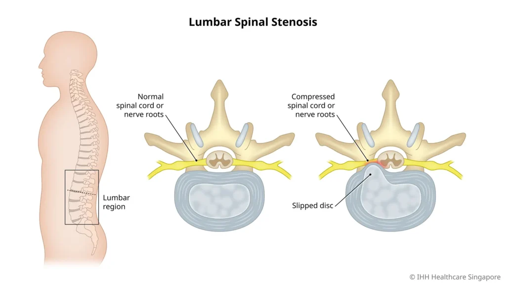
Lumbar spinal stenosis occurs when the spinal canal in the lower back narrows, reducing the space available for the spinal cord and nerve roots that pass through it. This narrowing compresses the nerves, producing the characteristic pain, cramping, and weakness associated with the condition. It can develop gradually through age-related degenerative changes or result from a combination of structural and lifestyle factors. Knowing what drives the narrowing, how it produces symptoms, and which non-surgical treatments offer the best evidence for relief is the foundation of effective long-term management.
What Happens Inside the Stenotic Spine
The lumbar spinal canal is a bony channel running through the vertebrae that houses and protects the lower spinal cord and nerve roots. In a healthy spine this canal provides ample space for neural structures to function without compression. In lumbar spinal stenosis, multiple structures can simultaneously encroach on this space — thickened ligaments bulging inward from behind, arthritic facet joints enlarging from the sides, and disc material protruding from the front — creating a three-dimensional narrowing that compresses the nerve roots and impairs their blood supply during upright posture and walking.
Causes and Risk Factors
Age-Related Degeneration
The most common cause of lumbar spinal stenosis is the gradual accumulation of degenerative changes in the spine that occur with normal aging. As spinal discs lose height and hydration over decades, the vertebrae move closer together, causing the facet joints at the back of each vertebral level to bear increased load. These joints respond by developing osteoarthritis — producing bone spurs and cartilage thickening that encroach on the spinal canal and lateral recesses where nerve roots exit. Simultaneously the ligamentum flavum — a flexible ligament running along the back of the spinal canal — thickens and loses elasticity with age, bulging inward and further narrowing the available space for neural structures.
Degenerative Disc Disease
As discussed in our guide on slipped disc herniated disc symptoms treatment and recovery, intervertebral discs naturally degenerate with age, losing height and structural integrity. This disc degeneration contributes directly to stenosis by allowing disc material to bulge posteriorly into the spinal canal and by reducing the vertical height of the neural foramina — the openings through which nerve roots exit the spine — compressing exiting nerve roots at multiple levels simultaneously.
Spondylolisthesis
Spondylolisthesis occurs when one vertebra slips forward over the one below it, distorting the spinal canal and compressing the nerve roots at that level. Degenerative spondylolisthesis — the most common form in older adults — results from facet joint arthritis weakening the posterior spinal stabilizers, allowing forward slippage typically at the L4-L5 level. This is one of the most common structural contributors to symptomatic lumbar stenosis in adults over 60.
Other Contributing Factors
- Congenital narrow canal: Some individuals are born with a naturally narrower spinal canal, meaning they develop symptomatic stenosis earlier in life and with less degenerative change than those with wider canals.
- Previous spinal surgery or injury: Scar tissue formation after spinal surgery, fractures, or trauma can contribute to canal narrowing and stenosis development over time.
- Obesity: Excess body weight accelerates lumbar disc degeneration and facet joint arthritis, hastening the degenerative cascade that produces stenosis. The relationship between lifestyle factors and spinal health is covered in our article on how everyday choices influence your body joint health.
Symptoms of Lumbar Spinal Stenosis
Neurogenic Claudication — The Hallmark Symptom
The most characteristic and diagnostically significant symptom of lumbar spinal stenosis is neurogenic claudication — a pattern of leg pain, heaviness, cramping, or weakness that develops after walking or standing for a period of time and relieves with sitting, bending forward, or lying down. Patients typically describe being able to walk only a limited distance — sometimes as little as half a block — before needing to stop and rest. Leaning forward on a shopping cart, walking uphill, or cycling are often better tolerated than walking on flat ground because forward flexion opens the spinal canal and temporarily increases space for compressed nerve roots.
Lower Back Pain
Most patients with lumbar spinal stenosis experience lower back pain alongside their leg symptoms, though the leg symptoms are often more disabling. The back pain is typically described as a dull, aching discomfort across the lower back that worsens with prolonged standing and extension and improves with sitting and flexion. Understanding the broader spectrum of back pain causes symptoms and risk factors provides important context for the lower back component of stenosis symptoms.
Leg Symptoms
- Bilateral leg symptoms: Unlike sciatica from a single-level disc herniation which typically affects one leg, lumbar stenosis often produces symptoms in both legs simultaneously, though one side is frequently worse than the other.
- Numbness and tingling: Pins-and-needles sensations in the thighs, calves, or feet are common and reflect compression of sensory nerve fibers within the narrowed canal.
- Leg weakness: Some patients notice progressive leg weakness during walking episodes, occasionally causing stumbling or foot drop in more severe cases.
Postural Dependence of Symptoms
The postural nature of stenosis symptoms is highly characteristic and clinically important. Symptoms consistently worsen with lumbar extension — standing upright, walking, or lying flat — because extension reduces the cross-sectional area of the spinal canal. Symptoms consistently improve with lumbar flexion — sitting, bending forward, or lying in the fetal position — because flexion opens the canal and temporarily decompresses the nerve roots. This flexion-relief pattern is what differentiates neurogenic claudication from vascular claudication, where leg pain from arterial insufficiency does not change with spinal position.
Diagnosis
Clinical Examination
An orthopedic or neurosurgical specialist evaluates gait, lower limb strength, sensation, and reflexes, and performs provocative tests assessing symptom reproduction with lumbar extension versus relief with flexion. The bicycle test — where symptoms develop with walking but not with stationary cycling in a flexed position — strongly supports neurogenic claudication from spinal stenosis over vascular causes.
Imaging
MRI is the definitive imaging investigation for lumbar spinal stenosis, providing precise visualization of canal dimensions, ligamentum flavum thickening, disc protrusions, facet joint hypertrophy, and nerve root compression at each spinal level. CT myelography is used when MRI is contraindicated or when surgical planning requires additional bony detail. X-rays assess spinal alignment, disc space narrowing, bone spur formation, and spondylolisthesis.
Non-Surgical Treatment Options
The encouraging evidence is that the majority of patients with lumbar spinal stenosis achieve meaningful symptom improvement with non-surgical treatment. Surgery produces faster initial relief but long-term outcomes at 4–10 years are comparable between surgical and well-managed conservative approaches for most patients.
Physical Therapy — The Foundation of Conservative Management
Physical therapy is the most evidence-based non-surgical treatment for lumbar spinal stenosis and should form the cornerstone of every conservative management program. A well-designed stenosis rehabilitation program focuses on several key components simultaneously.
Flexion-based exercises are central to stenosis rehabilitation because they open the spinal canal, decompress neural structures, and provide symptomatic relief while strengthening the muscles that maintain a flexed, canal-opening posture during daily activities. The Williams flexion exercise protocol — including knee-to-chest stretches, posterior pelvic tilts, and seated forward bending — is specifically designed for stenosis and produces meaningful pain reduction and functional improvement with consistent practice.
Core stabilization training strengthens the deep abdominal and lumbar stabilizer muscles that support the spine and reduce the dynamic canal narrowing that occurs with movement. A strong core reduces the degree of lumbar extension during walking and standing, directly addressing the positional mechanism that produces neurogenic claudication. Incorporating the habit changes discussed in our guide on how daily habits diet and remedies are effective for back pain alongside formal physical therapy significantly accelerates functional recovery.
Aquatic therapy is particularly valuable for stenosis patients because the buoyancy of water reduces spinal loading while allowing cardiovascular conditioning and strengthening exercises that would be too painful to perform on land. Many patients who can barely walk 50 meters on land can exercise comfortably for 30–45 minutes in a pool, maintaining fitness and building strength during the rehabilitation process.
Epidural Steroid Injections
Lumbar epidural steroid injections deliver corticosteroids directly into the epidural space surrounding compressed nerve roots, reducing inflammation and providing meaningful pain relief that typically lasts weeks to months. They are most valuable for patients with significant leg symptoms who cannot tolerate exercise due to pain, providing a window of reduced discomfort during which physical therapy can proceed more effectively. Transforaminal epidural injections — targeting the specific nerve root levels most compressed — tend to produce more precise and longer-lasting relief than interlaminar approaches for stenosis.
Medications
NSAIDs reduce inflammatory pain associated with facet joint arthritis and nerve root irritation, providing useful short-term symptomatic relief. Neuropathic pain medications including gabapentin and pregabalin address the nerve-related pain, numbness, and tingling that NSAIDs do not adequately control. Muscle relaxants help manage associated paraspinal muscle spasm. All medications should be used as adjuncts to active rehabilitation rather than as primary long-term treatments. Our guide on lifestyle medicine and its role in orthopedic pain explores the evidence for integrating lifestyle approaches alongside medical management.
Assistive Devices and Activity Modification
A walking stick or rollator walker encourages a slightly forward-flexed posture during walking, opening the spinal canal and increasing comfortable walking distance significantly for many stenosis patients. Activity modification — favoring cycling, swimming, and uphill walking over flat walking and standing — allows maintenance of cardiovascular fitness and functional independence while minimizing symptom provocation. Avoiding prolonged lumbar extension positions during daily activities, adjusting workstation ergonomics, and sleeping in a side-lying flexed position all reduce cumulative neural compression throughout the day.
Weight Management and Anti-Inflammatory Nutrition
Maintaining a healthy weight reduces mechanical load on degenerating lumbar discs and facet joints, slowing progression of the degenerative changes that drive stenosis worsening. Anti-inflammatory dietary patterns reduce systemic and local spinal inflammation that amplifies nerve root pain sensitivity. Our comprehensive guide on how changing your diet can help reduce pain naturally provides specific nutritional strategies directly applicable to stenosis management.
When to Consider Surgery
While this article focuses on non-surgical management, it is important to recognize when surgery becomes necessary. Surgical evaluation should be sought when conservative treatment has failed to provide adequate relief after 3–6 months, when walking distance is severely limited and significantly impairs quality of life, or when progressive neurological deficits develop including worsening leg weakness or loss of bladder and bowel control — the latter requiring emergency evaluation.
Conclusion
Lumbar spinal stenosis is a common, progressive but highly manageable condition that responds well to the right combination of physical therapy, activity modification, and appropriate medical treatment. The majority of patients achieve meaningful functional improvement and maintain good quality of life without surgery through consistent engagement with evidence-based conservative management. Early diagnosis, understanding your specific symptom pattern, and building the exercise and lifestyle habits that support spinal health give you the best possible foundation for long-term management of this condition.
Frequently Asked Questions
1. What is the main cause of lumbar spinal stenosis? The most common cause is age-related degenerative changes including facet joint arthritis, ligamentum flavum thickening, and disc degeneration that gradually narrow the spinal canal over decades.
2. Can lumbar spinal stenosis be reversed? The structural narrowing cannot be reversed without surgery, but symptoms can be very effectively managed and functional capacity significantly improved through physical therapy, exercise, and lifestyle modification.
3. What is the best exercise for lumbar spinal stenosis? Flexion-based exercises including knee-to-chest stretches, posterior pelvic tilts, and stationary cycling are most effective as they open the spinal canal and decompress nerve roots during activity.
4. How long can you live with spinal stenosis without surgery? Many people manage lumbar stenosis successfully with conservative treatment for years or decades. Surgery is not inevitable and is reserved for cases where conservative management has failed or significant neurological deficits develop.
5. Does walking worsen spinal stenosis? Prolonged walking on flat ground typically worsens stenosis symptoms due to lumbar extension. However, walking shorter distances within tolerance, using a walking aid, or walking uphill in a flexed posture is beneficial and should be encouraged.
6. What makes spinal stenosis symptoms worse? Prolonged standing, walking on flat surfaces, lumbar extension exercises, lying flat, and downhill walking consistently worsen symptoms by reducing spinal canal cross-sectional area.
7. Is cycling safe with lumbar spinal stenosis? Yes — stationary and road cycling in a forward-flexed position is one of the best-tolerated exercises for stenosis patients, opening the spinal canal and maintaining cardiovascular fitness without provoking neurogenic claudication.
8. Can stenosis cause permanent nerve damage? Prolonged severe nerve compression can cause lasting neurological deficits. Progressive weakness, worsening numbness, or loss of bladder and bowel control are warning signs requiring prompt surgical evaluation.
9. Does losing weight help spinal stenosis? Yes — weight loss reduces mechanical load on degenerating spinal structures, decreases inflammatory drive, and improves physical therapy outcomes significantly in overweight patients with stenosis.
10. What is the difference between sciatica and spinal stenosis? Sciatica typically affects one leg and often results from a single herniated disc compressing one nerve root. Spinal stenosis more commonly affects both legs, worsens specifically with walking and standing, and relieves with sitting and forward bending.
Disclaimer
The information provided in this article is for educational and informational purposes only and does not constitute professional medical advice, diagnosis, or treatment.
This article is for education purpose only and is not a diagnosis or a substitute for medical advice.